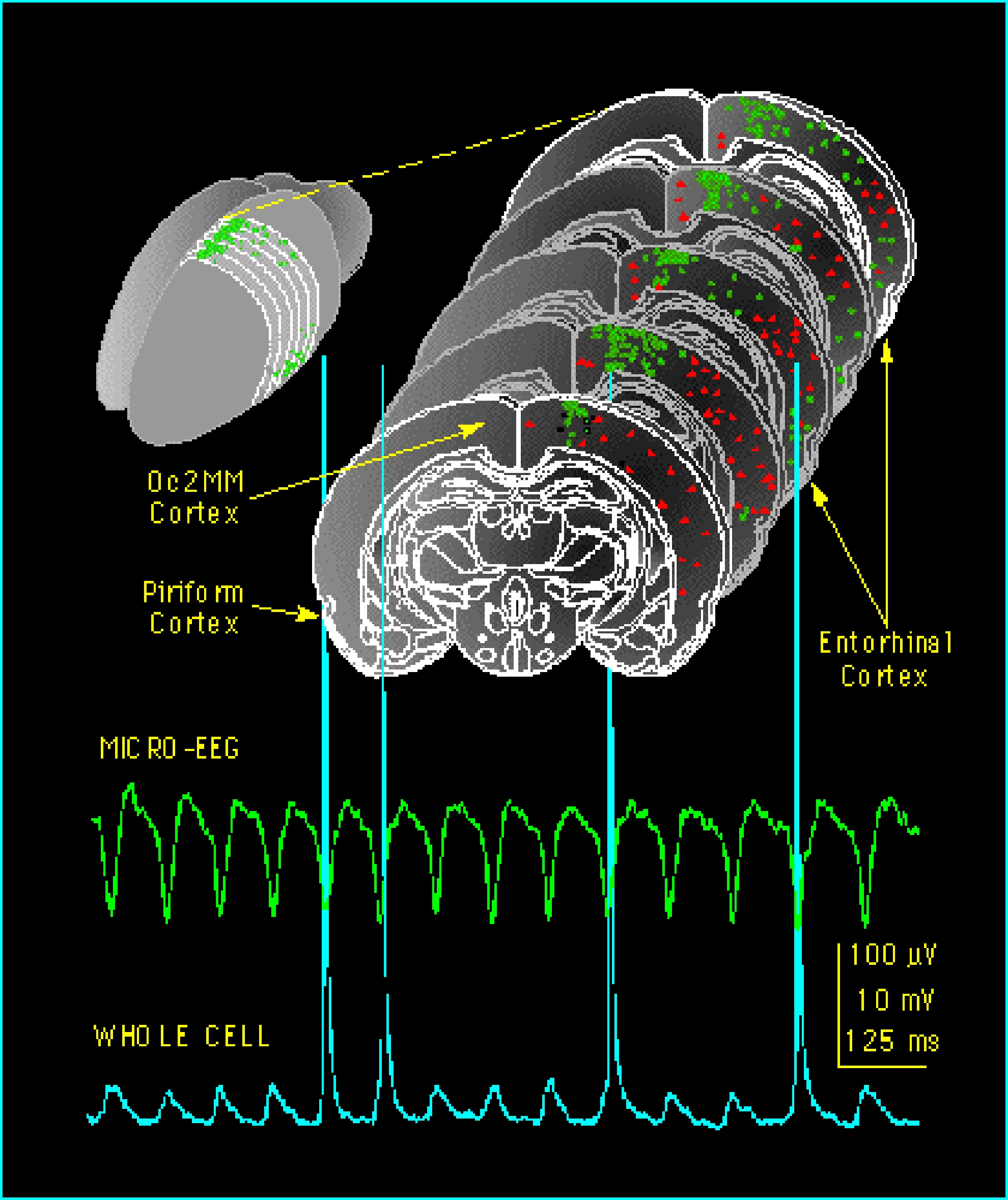
Micro-EEG
It represents locations in rat brain slices where theta EEG-like activity can be recorded (green dots) and where this activity was not evident (red dots), together with recordings of theta field 'micro-EEG' and single neuron membrane potentials.

