NEURONAL DIVERGENCE
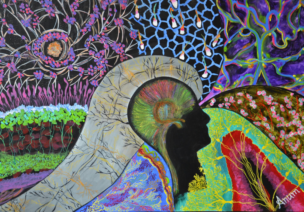
The painting depicts the versatility and sophistication of neuroscience. As an independent artist and medical doctor I am fascinated by artistic expression of scientific excellence. It is a 100x70x4 cm acrylic canvas painting portraying an array of images demonstrating the unique power of art to convey the incredible beauty and complexity of the brain. In the right lower corner we see a transverse section of cerebellar lobe with radiating purkinje cells (yellow). Above it is the image of olfactory bulb, a neuronal structure in vertebrate forebrain involved in sense of smell. The right upper corner shows the midbrain vasculature of zebrafish larvae. Blood (blue) is seen traveling along endothelial cells (yellow-green). Next, to the left is the sensory epithelium of mouse utricle (inner ear). In the upper left corner there is an image of brain cancer cells being used currently in studying neuronal development by researchers. Below this the image of various layers of retina can be appreciated. The various cell types in each layer example cones (pink), rods(green) can also be visualized. Further below in grey is a stylized section of cerebral cortex showing network of branching pyramidal cells. Next to this is the image of retina painted with all its intricate branching vasculature. All these magnificent images converge at the center where a human figure is showcasing the brain connectome.

