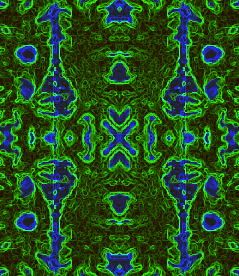
abstract_Lesion Matrix_by_R. Matthew Hutchison
Multiple sclerosis is characterized by the demyelination of neurons that, at a large-scale, can be viewed on an MRI image as white matter intensity changes. Critical to analysis is the proper identification and boundary detection of lesions so that their progress can be tracked over time and in response to treatment. The image (a mosaic of a horizontal section of an MS patient T2 MRI image) highlights the computational challenge of automatic lesion detection and the need to consider the underlying neuroanatomy and image properties.

