abstract_Anatomical Patterns of Brain Tissue Clusters Segmented Individually in the RGB (Red, Green, Blue) Color Components_by_Peifang Guo
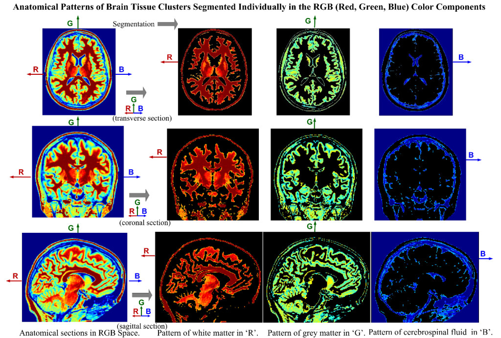
This image illustrates that three labeled classes of white matter (WM), grey matter (GM) and cerebrospinal fluid (CSF) in three anatomical sections of T1w 3D MRI (transverse, coronal and sagittal sections) are mapped into the RGB (red, green, blue) color space; then using the k-means clustering approach, the three clusters of WM, GM and CSF patterns in the three anatomical brain sections are segmented graphically in an automatic fashion in different components of ‘R’ (with the pattern of WM), the ‘G’ (with the pattern of GM) and the ‘B’ (with the pattern of CSF) individually.

